Acoustic Neuroma Audiogram: Diagnosis Guide 2024
An acoustic neuroma audiogram serves as a crucial diagnostic tool for detecting vestibular schwannoma, revealing characteristic hearing loss patterns that help physicians identify this benign brain tumor. This comprehensive hearing test can detect the asymmetric sensorineural hearing loss typically associated with acoustic neuromas, making it an essential first step in the diagnostic process for patients experiencing unilateral hearing symptoms.
Understanding Acoustic Neuroma and Audiogram Testing
An acoustic neuroma, medically known as vestibular schwannoma, is a non-cancerous tumor that develops on the eighth cranial nerve connecting your inner ear to your brain. This slow-growing tumor affects approximately 1 in 100,000 people annually in the United States, with most cases diagnosed between ages 30-60. The condition primarily impacts the nerve responsible for hearing and balance, making audiological testing crucial for early detection.
The audiogram represents the gold standard initial screening tool for acoustic neuroma diagnosis. This painless hearing test measures your ability to hear sounds at different frequencies and volumes, creating a visual map of your hearing capabilities. Healthcare providers specifically look for asymmetric sensorineural hearing loss, which occurs in approximately 95% of acoustic neuroma patients according to 2024 medical research.
Characteristic Hearing Loss Patterns in Acoustic Neuroma
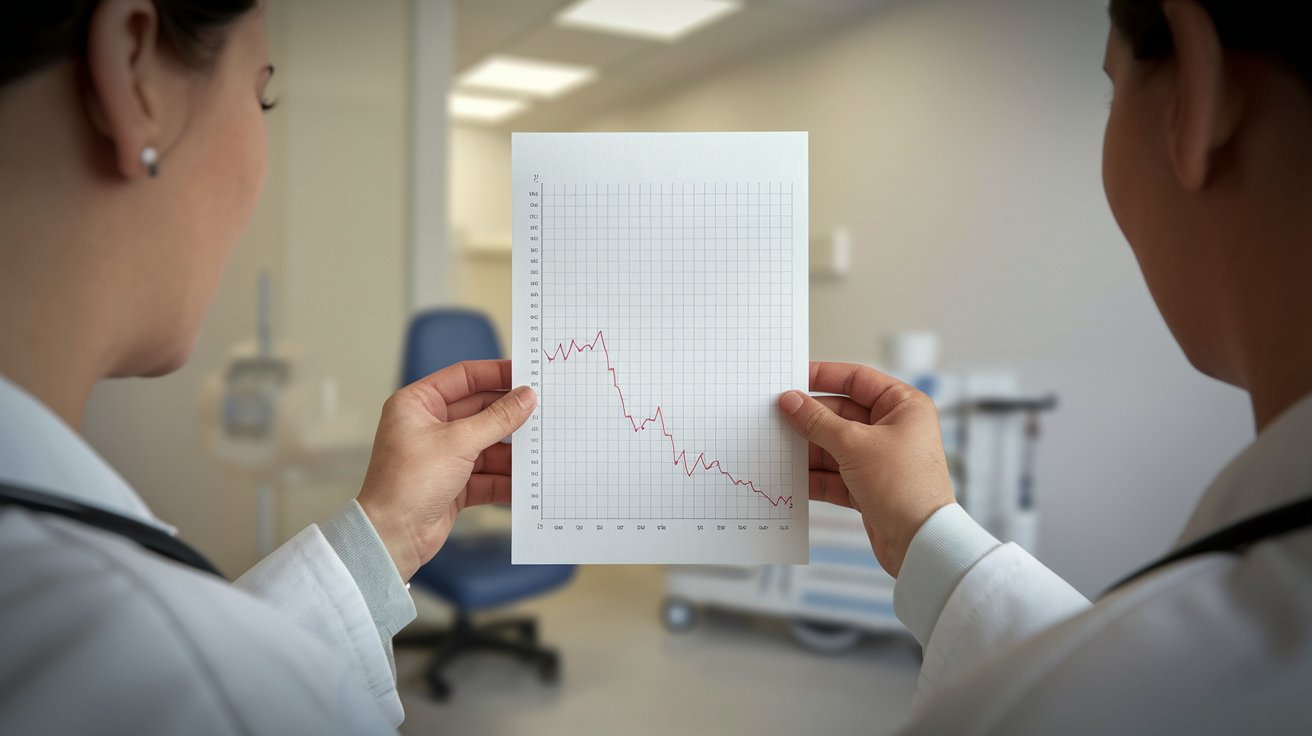
The acoustic neuroma hearing loss pattern displays distinctive characteristics that differentiate it from other hearing conditions. Patients typically experience progressive, unilateral sensorineural hearing loss that worsens over time. The hearing loss often begins in high frequencies and gradually affects lower frequencies, creating a distinctive downward slope on the audiogram chart.
Research from 2024 indicates that asymmetric hearing loss greater than 15 decibels between ears should prompt further investigation for acoustic neuroma. The tumor’s pressure on the auditory nerve creates this characteristic pattern, with some patients experiencing sudden hearing loss episodes. Word recognition scores also deteriorate disproportionately compared to pure tone thresholds, a phenomenon known as retrocochlear hearing loss.
High-Frequency Hearing Loss Indicators
High-frequency hearing loss represents the earliest and most common audiometric finding in acoustic neuroma patients. The tumor’s compression of nerve fibers responsible for high-pitch sound transmission creates characteristic drops in hearing sensitivity above 4000 Hz. Audiologists specifically monitor for hearing thresholds exceeding 25 decibels in frequencies between 4000-8000 Hz, as these ranges show the most significant deterioration in early-stage acoustic neuromas.
Word Recognition Score Decline
Patients with acoustic neuroma demonstrate disproportionately poor word recognition scores compared to their pure tone thresholds, a hallmark of retrocochlear pathology. While their ability to hear tones may remain relatively intact, their capacity to understand speech becomes significantly impaired. This disconnect between hearing sensitivity and speech comprehension serves as a red flag for audiologists, often prompting referral for additional testing including MRI imaging.
Acoustic Neuroma Symptoms Beyond Hearing Loss
Acoustic neuroma symptoms extend beyond hearing impairment, encompassing a range of neurological manifestations that develop as the tumor grows. Unilateral tinnitus affects approximately 83% of patients, presenting as ringing, buzzing, or roaring sounds in the affected ear. Balance problems and dizziness occur in about 61% of cases, ranging from mild unsteadiness to severe vertigo episodes that significantly impact daily activities.
Facial numbness and weakness represent more advanced acoustic neuroma symptoms that occur when larger tumors compress the facial nerve. Some patients experience ear fullness or pressure sensations, similar to having water trapped in the ear. Headaches, particularly those localized to one side, may develop as the tumor increases intracranial pressure, affecting approximately 32% of patients with larger acoustic neuromas.
Balance and Vestibular Symptoms
Balance problems associated with acoustic neuroma result from tumor pressure on the vestibular portion of the eighth cranial nerve. Patients often report feeling unsteady when walking, particularly in dark environments or on uneven surfaces. The gradual onset allows many individuals to compensate through visual and proprioceptive cues, making these symptoms less obvious than the hearing loss component.
Tinnitus and Ear-Related Symptoms
Unilateral tinnitus serves as one of the most reliable early warning signs of acoustic neuroma, occurring in the vast majority of cases. The phantom sounds typically remain constant rather than pulsatile, distinguishing them from vascular causes. Patients describe various sound qualities including high-pitched ringing, buzzing, hissing, or roaring noises that persist throughout the day and often worsen in quiet environments.
Advanced Diagnostic Methods Beyond Audiogram
While the audiogram serves as the initial screening tool, definitive acoustic neuroma diagnosis requires advanced imaging studies. Magnetic resonance imaging (MRI) with gadolinium contrast remains the gold standard for acoustic neuroma detection, capable of identifying tumors as small as 1-2 millimeters. The 2024 acoustic neuroma MRI protocol includes high-resolution T1-weighted sequences that provide detailed visualization of the internal auditory canal and cerebellopontine angle.
Additional audiological tests complement the standard audiogram, including acoustic reflex testing, otoacoustic emissions, and auditory brainstem response (ABR) testing. These specialized evaluations help differentiate between cochlear and retrocochlear hearing loss, providing crucial information about nerve function integrity. ABR testing shows particular value in detecting acoustic neuromas, with abnormal results occurring in approximately 90% of cases.
MRI Imaging Protocols and Accuracy
The acoustic neuroma MRI protocol utilizes thin-slice imaging through the internal auditory canals, typically 1-2 millimeter sections with T1-weighted sequences after gadolinium administration. Modern 3-Tesla MRI machines achieve diagnostic accuracy rates exceeding 99% for acoustic neuroma detection. Radiologists specifically examine the cerebellopontine angle and internal auditory canal for enhancing masses that appear bright on contrast-enhanced images.
Auditory Brainstem Response Testing
Auditory brainstem response testing measures electrical activity along the auditory pathway from the ear to the brainstem, providing objective assessment of nerve function. ABR abnormalities in acoustic neuroma patients include prolonged wave V latencies and increased interaural wave V latency differences. This test proves particularly valuable when MRI is contraindicated or when screening high-risk patients with family histories of acoustic neuroma.
Treatment Options and Surgical Considerations
Acoustic neuroma treatment depends on multiple factors including tumor size, growth rate, patient age, and hearing status. Small tumors under 1.5 centimeters may be monitored with serial MRI scans every 6-12 months, as approximately 30% show no significant growth over five years. Active surveillance allows patients to maintain quality of life while avoiding treatment-related risks, particularly appropriate for elderly patients or those with significant comorbidities.
Acoustic neuroma surgery becomes necessary for larger tumors or those showing rapid growth, with three primary surgical approaches available. The translabyrinthine approach provides excellent tumor visualization but sacrifices hearing, while the retrosigmoid approach attempts hearing preservation in suitable candidates. Stereotactic radiosurgery using Gamma Knife or CyberKnife technology offers a non-invasive alternative for tumors under 3 centimeters, achieving tumor control rates exceeding 90% in recent studies.
Surgical Approaches and Outcomes
Modern acoustic neuroma surgery techniques focus on maximizing tumor removal while preserving facial nerve function and, when possible, hearing. The middle fossa approach allows hearing preservation attempts in patients with good preoperative hearing and small tumors confined to the internal auditory canal. Surgical outcomes in 2024 show facial nerve preservation rates exceeding 95% for experienced surgeons, with hearing preservation possible in 30-70% of appropriate candidates.
Radiation Therapy and Stereotactic Options
Stereotactic radiosurgery delivers precisely targeted radiation to the acoustic neuroma while minimizing exposure to surrounding healthy tissue. This outpatient procedure requires no incision and allows most patients to return to normal activities within days. Long-term tumor control rates approach 95% at 10 years, making it an attractive option for patients seeking to avoid traditional surgery while maintaining current hearing levels.
Prognosis and Long-term Management
The acoustic neuroma prognosis remains generally favorable given the benign nature of these tumors, with five-year survival rates approaching 100%. However, treatment-related complications can significantly impact quality of life, including permanent hearing loss, facial weakness, and balance problems. Modern treatment approaches prioritize functional outcomes, with multidisciplinary teams including neurotologists, neurosurgeons, and radiation oncologists collaborating to optimize individual patient care.
Long-term follow-up care involves regular audiogram monitoring, balance assessments, and periodic MRI scans to detect recurrence or growth in residual tumor tissue. Patients require ongoing support for hearing rehabilitation, including hearing aid fitting or cochlear implant evaluation for those with significant hearing loss. Physical therapy helps address balance issues, while facial rehabilitation programs assist patients experiencing facial nerve complications from treatment.
Related video about acoustic neuroma audiogram
This video complements the article information with a practical visual demonstration.
Most asked questions about acoustic neuroma audiogram
Can an audiogram detect acoustic neuroma?
An audiogram can identify characteristic hearing loss patterns suggestive of acoustic neuroma, particularly asymmetric sensorineural hearing loss and poor word recognition scores. However, definitive diagnosis requires MRI imaging, as the audiogram serves as a screening tool that indicates the need for further testing rather than providing a conclusive diagnosis.
What is the hallmark of acoustic neuroma on audiogram?
The hallmark of acoustic neuroma on audiogram is asymmetric sensorineural hearing loss, typically affecting one ear more than the other. Patients show disproportionately poor word recognition scores compared to pure tone thresholds, high-frequency hearing loss, and hearing differences greater than 15 decibels between ears.
What is vestibular schwannoma audiogram pattern?
Vestibular schwannoma audiogram patterns show progressive unilateral sensorineural hearing loss with a characteristic downward slope affecting high frequencies first. The pattern includes asymmetric hearing between ears, reduced word discrimination scores, and retrocochlear hearing loss characteristics that distinguish it from inner ear disorders.
How accurate is audiogram testing for acoustic neuroma?
Audiogram testing shows high sensitivity for detecting hearing changes associated with acoustic neuroma, identifying characteristic patterns in approximately 95% of cases. However, specificity is lower as other conditions can cause similar hearing loss patterns, making MRI imaging necessary for definitive diagnosis and tumor visualization.
What are red flags for acoustic neuroma on hearing tests?
Red flags for acoustic neuroma on hearing tests include unilateral hearing loss, asymmetric hearing between ears exceeding 15 decibels, poor word recognition scores disproportionate to pure tone loss, high-frequency hearing loss progression, and abnormal acoustic reflex patterns. These findings warrant immediate referral for MRI evaluation.
Should I get regular audiograms if I have acoustic neuroma?
Yes, regular audiograms are essential for acoustic neuroma monitoring, typically performed every 6-12 months during active surveillance or annually after treatment. These tests track hearing changes over time, guide treatment decisions, and help optimize hearing rehabilitation strategies including hearing aid fitting or cochlear implant evaluation.
| Diagnostic Aspect | Key Features | Clinical Significance |
|---|---|---|
| Audiogram Pattern | Asymmetric sensorineural hearing loss >15dB | Primary screening indicator |
| Word Recognition | Disproportionately poor scores | Suggests retrocochlear pathology |
| Frequency Impact | High-frequency loss predominant | Early detection opportunity |
| MRI Confirmation | 99% diagnostic accuracy | Definitive diagnosis and treatment planning |